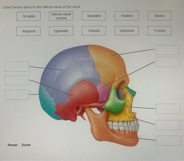
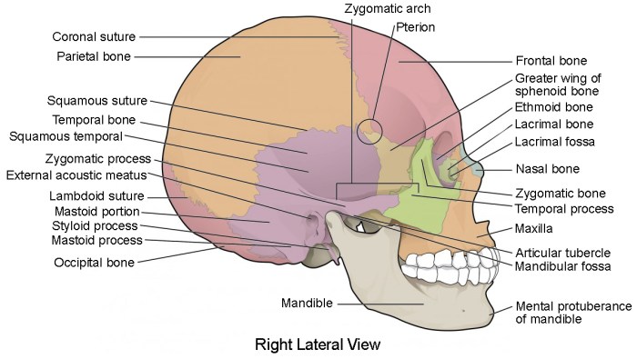
Label bones seen in the lateral view of the skull – Labeling the bones seen in the lateral view of the skull is a crucial step in understanding the anatomy of the human skull. This comprehensive guide will provide an in-depth overview of the bones visible in this perspective, their location, shape, landmarks, and functions.
Beginning with the frontal bone, we will explore its prominent position at the forehead, its role in forming the anterior cranial fossa, and the various landmarks it bears, including the supraorbital margin and the frontal sinuses. Moving posteriorly, we will examine the parietal bone, its contribution to the calvaria, and the parietal foramina that transmit blood vessels and nerves.
Label Bones Seen in the Lateral View of the Skull

The lateral view of the skull presents a comprehensive perspective of the cranial bones that constitute the protective structure encasing the brain. This article will delve into the specific bones visible in this view, exploring their location, shape, landmarks, and functions.
Frontal Bone
Location: Anterior aspect of the skull
Shape: Convex and dome-shaped
Landmarks:
- Frontal sinus
- Glabella
- Supraorbital notch
Functions:
- Protects the frontal lobe of the brain
- Provides attachment points for facial muscles
- Contributes to the formation of the forehead and orbits
Parietal Bone
Location: Superolateral aspect of the skull
Shape: Quadrilateral with curved borders
Landmarks:
- Parietal eminence
- Sagittal suture
- Temporal line
Functions:
- Protects the parietal lobe of the brain
- Provides attachment points for muscles of the scalp
- Contributes to the formation of the cranial vault
Temporal Bone, Label bones seen in the lateral view of the skull
Location: Lateral aspect of the skull
Shape: Complex and irregular
Landmarks:
- Mastoid process
- Styloid process
- External auditory meatus
Functions:
- Protects the temporal lobe of the brain
- Houses the organs of hearing and balance
- Provides attachment points for muscles of mastication
Occipital Bone
Location: Posterior aspect of the skull
Shape: Trapezoidal
Landmarks:
- Foramen magnum
- Occipital condyles
- External occipital protuberance
Functions:
- Protects the occipital lobe of the brain
- Forms the base of the skull
- Provides attachment points for muscles of the neck
Sphenoid Bone
Location: Central and anterior aspect of the skull
Shape: Bat-shaped with a complex structure
Landmarks:
- Sella turcica
- Optic canal
- Pterygoid process
Functions:
- Forms the base of the skull and the floor of the middle cranial fossa
- Protects the pituitary gland and optic nerves
- Provides attachment points for muscles of the eye and jaw
Ethmoid Bone
Location: Anterior and superior aspect of the skull
Shape: Labyrinthine and delicate
Landmarks:
- Cribriform plate
- Lateral mass
- Conchae
Functions:
- Forms the roof of the nasal cavity and part of the orbit
- Supports the olfactory bulb
- Contributes to the formation of the paranasal sinuses
FAQ: Label Bones Seen In The Lateral View Of The Skull
What is the largest bone in the lateral view of the skull?
The parietal bone is the largest bone in the lateral view of the skull.
What is the function of the temporal bone?
The temporal bone houses the organs of hearing and balance, and also contains the mandibular fossa, which articulates with the mandible.

